Table of Contents
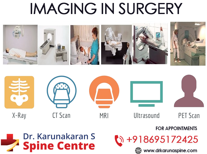
Imaging in surgery is very important. The prevalent imaging tests include X-rays, CT scan, MRI, mammogram, ultrasound, fluoroscopy and PET scans. While X-rays and CT scans employ ionizing radiation, MRI and ultrasound use magnetic and sound waves respectively. PET scans use radio tracers.
CT scan is advantageous over MRI for imaging calcified tissues and provides more information about osteoarthritis and fractures. MRI is better than CT scan to study water containing tissues and to detect abnormalities like bulging disc, disc herniation, pinched nerves and soft tissue problems. In cases like those of pregnant women where X-rays are contraindicated, MRI are used for imaging. However if metallic implants are present, MRI cannot be used.
MRI detects spine alignment, birth defects in the vertebrae, trauma injury in the bone or discs, compression or inflammation of spinal cord nerves, infection or tumors in vertebrae or spinal cord.
Computed Tomography or CT scan brings together the capacity of X ray and computers to give 360 degree cross sectional view of the bony structure of spine vertebrae with optimum accuracy.